

辛伐他汀透過保護粒線體對抗血管緊張素II所引發的心臟衰竭
在常見的心血管疾病包括「心臟衰竭」,通常伴隨著有粒線體功能障礙的情形。而羥甲基戊二酸單醯輔酶A還原酶(他汀類藥物)能夠抑制有害的氧自由基的產生,因此在慢性心臟衰竭中具有心臟保護作用。但是,他汀類藥物在心臟衰竭過程中是如何達到效果的? 以及對於心肌細胞內的能量提供的來源「粒線體」是否也有保護作用?這是我們研究團隊欲探索的項目。
透過心衰竭大鼠的動物實驗方式,我們團隊將大鼠分成: 1) 給予血管緊張素II(1.5 mg / kg /天/14天。2)予血管緊張素II合併給予辛伐他汀組(口服,10 mg/kg)經治療14天後在接下來的14天中停止給藥,終止實驗。
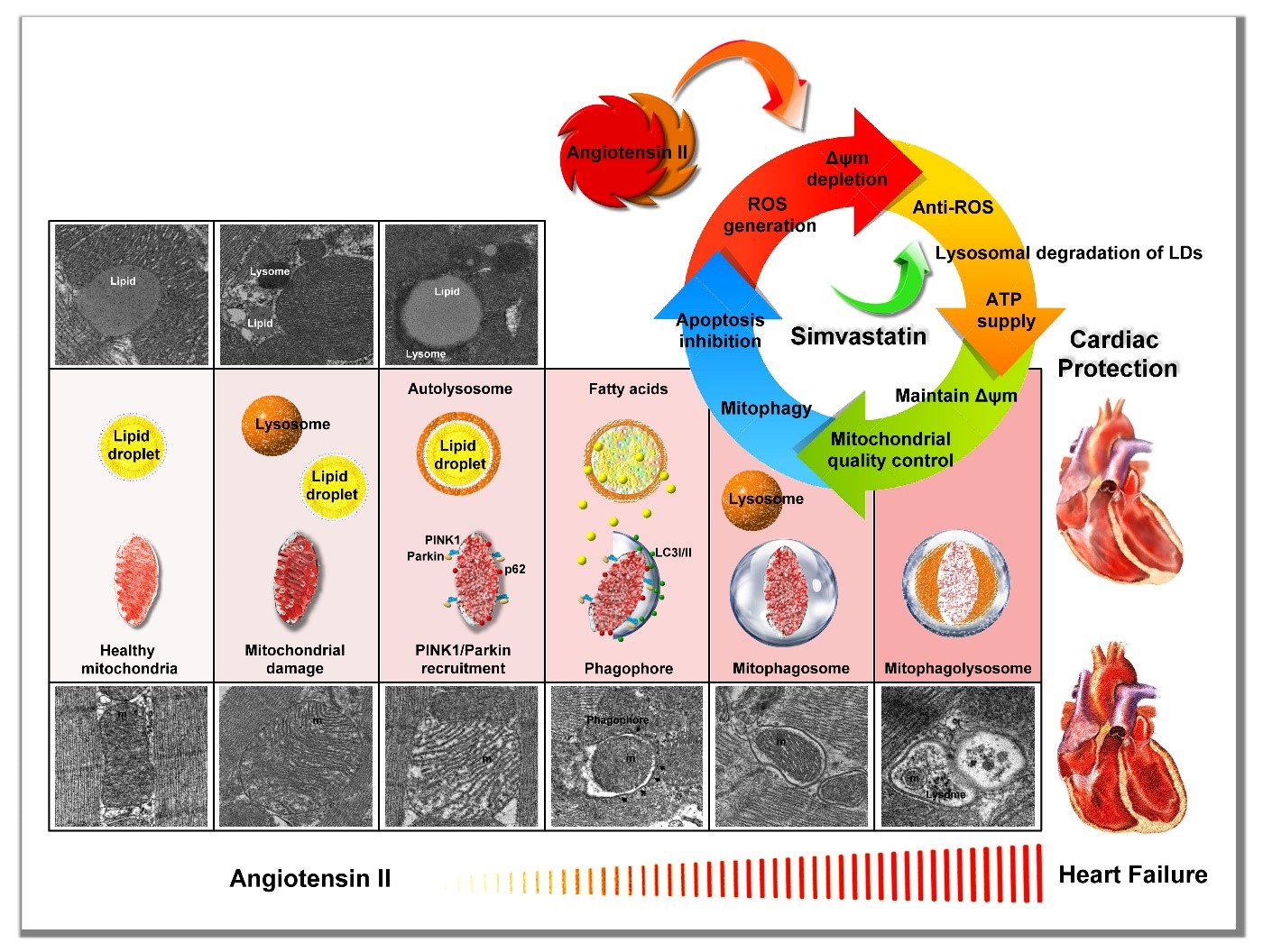
我們的研究成果發現:血管緊張素II會促進細胞內氧自由基的生成進而降低了粒線體的膜電位,導致粒線體的失能或是被破壞。然而透過給予辛伐他汀能夠有效地避免胞內氧自由基的生成進而維持了粒線體的膜電位使粒線體不致失能。而其中的關鍵在於辛伐他汀能夠在粒線體受損透過溶酶體將脂質油滴分解成脂肪酸產生能量,讓細胞有機會透過細胞自噬體來將受損的粒線體修復、或是移除。進而保護並維持細線體的功能,減緩心臟衰竭過程中對於心肌細胞的傷害。
圖形摘要
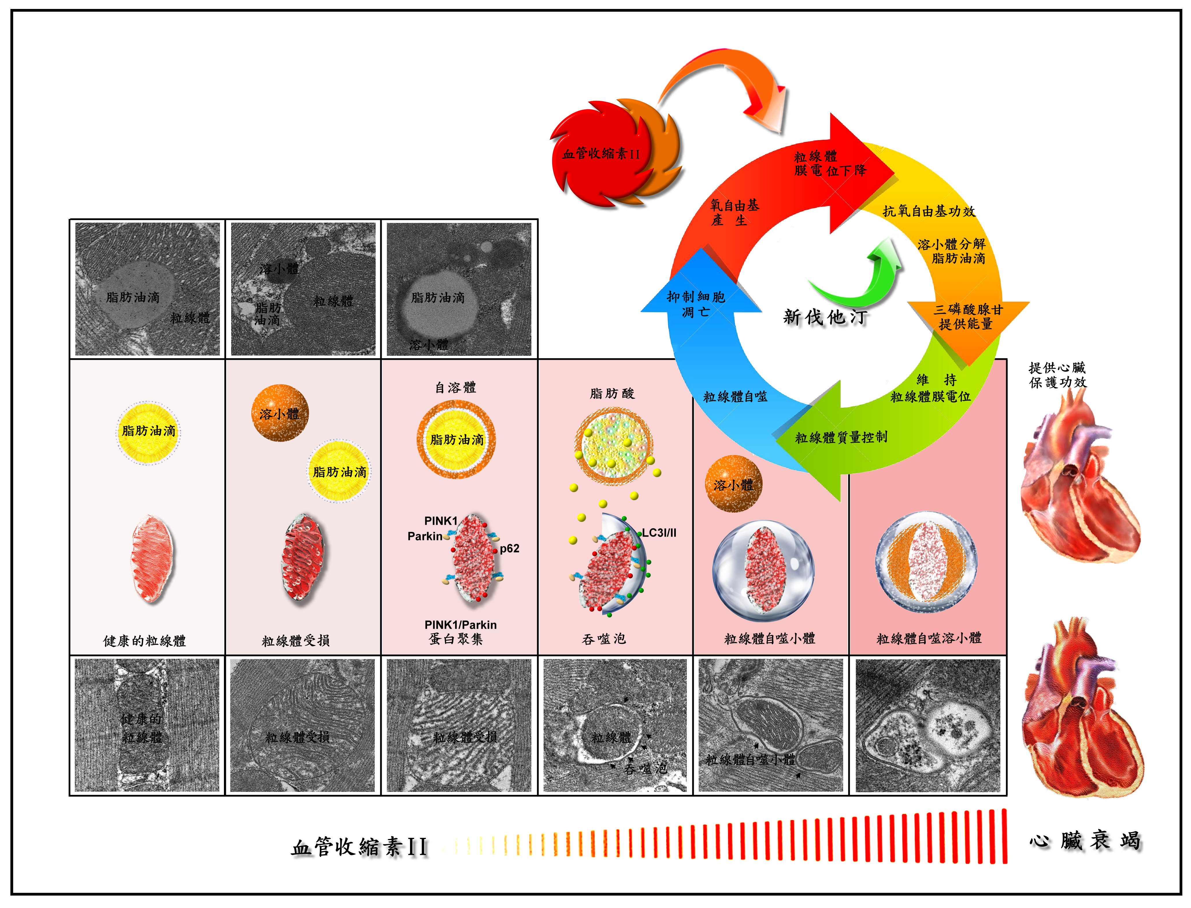
圖片說明: 血管緊張素II促進細胞內氧自由基的增加,降低了粒線體的膜電位,促使粒線體產生障礙無法提供心肌能量。透過給予辛伐他汀能夠有效地降低氧自由基並維持了粒線體的膜電位。同時辛伐他汀促進脂質油滴聚集於受損的粒腺體周圍並分解成脂肪酸提供粒線體透過自噬體的方式來自我修復,或是將無用的粒線體移除、進而維持細線體的功能減緩心臟衰竭對於心肌所帶來的傷害。
本篇為高雄醫學大學2019年月傑出論文10月份得獎文章,代表作者為醫學院呼吸治療學系劉博侖教授。
本校主要研究者之簡介:
論文第一作者為本校臨床醫學所博士班學生謝炯昭主治醫師,其為本校呼吸治療學系劉博侖教授所領導的團隊的一員。感謝科技部提供研究經費。
研究聯繫Email:
期刊出處:
British
Journal of Pharmacology. 2019
Oct;176(19):3791-3804.
期刊線上參閱網址:
Mitochondrial protection by
simvastatin against Angiotensin II-mediated heart failure
BACKGROUND AND PURPOSE
Mitochondrial dysfunction
plays a role in the progression of cardiovascular diseases including heart
failure. 3-Hydroxy-3-methylglutaryl-coenzyme A reductase inhibitors (statins),
which inhibit reactive oxygen species (ROS) synthesis, show cardioprotective
effects in chronic heart failure. However, the beneficial role of statins in
mitochondrial protection in heart failure remains unclear.
EXPERIMENTAL APPROACH
Rats
were treated with Angiotensin II (Ang II) (1.5 mg/kg/day) or co-administered
simvastatin (oral, 10 mg/kg) for 14 days; and then administration was stopped
for the following 14 days. Cardiac structure/function was examined by wheat
germ agglutinin staining and echocardiography. Mitochondrial morphology and the
numbers of lipid droplets, lysosomes, autophagosomes, and mitophagosomes were
determined by transmission electron microscopy. Human cardiomyocytes were
stimulated and intracellular ROS and mitochondrial membrane potential (ΔΨm)
changes were measured by flow cytometry and JC-1 staining, respectively.
Autophagy and mitophagy-related and mitochondria-regulated apoptotic proteins
were identified by immunohistochemistry and western blotting.
Graphical Abstract
This
article-“Mitochondrial protection by simvastatin against angiotensin
II-mediated heart failure” , written by Rept. Author Professor Po-Len Liu
from Department of
Respiratory Therapy in College of Medicine,,
is presented for Kaohsiung Medical University 2019 Monthly Excellent Paper
Award in Oct.
Main researcher Intro.
Summary
scheme of the mitochondrial protection mechanism of simvastatin in Ang
II-induced HF. Simvastatin might reduce ROS generation, regulate LDs and
lysosome levels to provide energy to maintain ΔΨm, regulate
mitochondrial quality control to promote mitophagy, and prevent
mitochondrial-regulated apoptosis.
Researcher
The first author of the paper is the attending
physician Chong-Chao Hsieh, a student of the School of Clinical Medicine,
and a member of the team led by Professor Po-Len Liu of the Department of
Respiratory Therapy, College of Medicine, Kaohsiung Medical University. Thanks
to the Ministry of Science and Technology for providing research funding.
Author Email
Paper cited from:
Paper online website
擾亂登革病毒增殖的殺手:前列腺素(Prostasin)基因
登革病毒是藉由蚊子傳播的病毒,埃及斑蚊(Aedes aegypti)或白線斑(Aedes albopictus)是最主要的病媒蚊,登革病毒感染會導致威脅生命的疾病,例如登革出血熱或登革熱休克症候群。在熱帶和亞熱帶地區是一個嚴重的公共衛生問題,台灣位於熱帶和亞熱帶地區的交界處,每年都有登革病毒感染病例,特別是在台灣南部,在2014年至2015年,台南和高雄就曾經爆發登革病毒感染,感染病毒的人數分別超過一萬和兩萬多人,且造成百餘人死亡。目前並尚未有有效的疫苗和藥物用於預防和臨床治療,因此,尋找具有抗登革病毒感染之潛力藥物標靶是重要的研究議題。眾所周知,病毒是一種寄生的病原體,也就是說,病毒的增殖需要依賴宿主基因表達的支持。在過去數年中,高雄醫學大學李景欽教授和陳彥旭醫師合作致力於了解登革病毒複製機制並尋找引起登革熱疾病的標靶基因。根據臨床檢驗分析,研究團隊首先發現,相較於健康個體,登革熱患者血液樣本中前列腺素(prostasin) (一個細胞膜蛋白酶)的表達量較低,並進而揭示前列腺素表達量跟登革病毒RNA感染量呈現負相關,意即,病毒感染量越多,前列腺素量越少。而回到實驗室進行一系列的細胞學實驗,結果顯示登革病毒感染確實會顯著降低前列腺素基因表現量。因此,研究小組認為登革病毒感染會抑制前列腺素表現的原因是因為前列腺素會對病毒登革病毒的增殖產生干擾作用。為了證明這個假設,研究小組在登革病毒感染的細胞或小鼠中大量表達外源性的前列腺素,結果發現大量表達的前列腺素確實可以抑制病毒增殖並保護小鼠免於受登革病毒感染的死亡威脅,意即減緩老鼠的死亡率。由進一步的抑制機轉的研究顯示,前列腺素對登革病毒增殖的抑制作用是藉由抑制上皮生長因子受體(epithelial growth factor receptor; EGFR)的表達,進而導致Akt /NF-B調控的環氧合酶-2(cyclooxygenase-2; COX-2)訊息路徑受到抑制。先前研究小組的研究已經證實登革病毒增殖需要COX-2訊息路徑的活化。同時根據報導,EGFR活化路徑會擾亂細胞干擾素(Interferon)的抗病毒反應,進而達到幫助病毒增殖的目的。因此,前列腺素透過降低EGFR的表達,進而恢復干擾素和干擾素所誘發的抗病毒基因的表達,包括2'-5'-寡腺苷酸合成酶1 (OAS1),OAS2,OAS3和雙鏈RNA依賴性蛋白激酶(PKR)而達到抗病毒目的。總之,我們的結果證明前列腺素的表達是一個值得做為臨床診斷標記的特徵,同時也提供治療登革病毒感染的潛在靶標。
圖形摘要
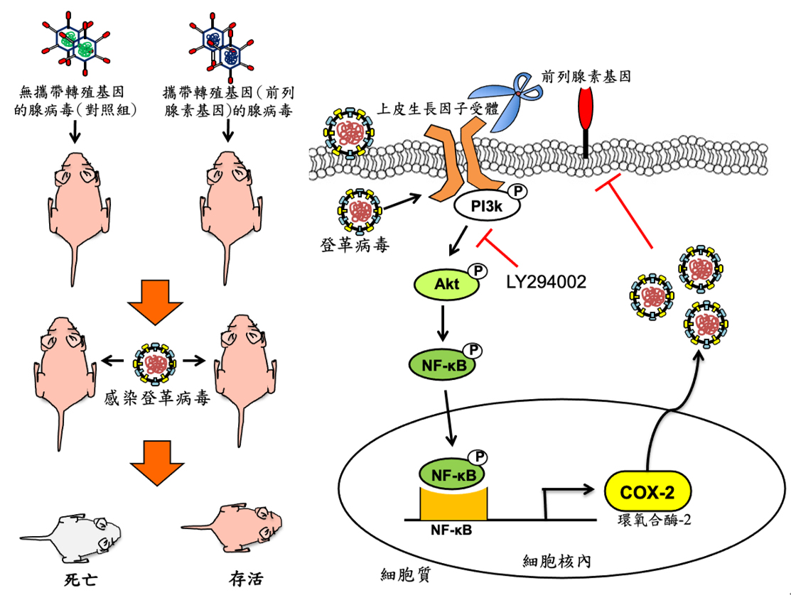
前列腺素藉由降低EGFR蛋白進而抑制登革病毒複製的模型。
1. 前列腺素的外源表達保護老鼠免受登革病毒感染造成的的死亡並抑制病毒的繁殖。
2. 前列腺素的抗病毒機制是降低上皮生長因子受體(EGFR)的表達量,導致抑制Akt/NF-B所調控的環氧合酶2(COX-2)訊息傳遞路徑而不利於病毒增殖。
本校主要研究者之簡介:
團隊負責人是本校生物科技學系李景欽教授,合作的臨床檢驗負責人是陳彥旭醫師/教授。 合著者包括:林俊光(第一作者,博士生),曾敬凱,吳宇軒,林俊佑(臨床醫生; 博士),黃崇豪(臨床醫師; 博士),王文宏博士,廖志中教授。
研究聯繫Email:
李景欽: jclee@kmu.edu.tw
陳彥旭: infchen@gmail.com
期刊出處:
The Journal of Infectious Diseases, the 16th of April 2019. “Prostasin impairs epithelial growth factor activation to suppress dengue virus propagation”
期刊線上參閱網址:
https://academic.oup.com/jid/article-abstract/219/9/1377/5200723?redirectedFrom=fulltext
Prostasin gene expression interferes dengue virus propagation
Dengue virus (DENV) is a mosquito-transmitted virus. The most important vector mosquito is the Aedes aegypti or Aedes albopictus. DENV infection leads to life-threatening diseases, such as dengue hemorrhagic fever or dengue shock syndrome. It is a serious public health problem in tropical and subtropical regions of the world. Taiwan is located at the junction of tropical and subtropical regions and had cases of dengue infections every year, especially in southern Taiwan. An outbreak of dengue infections occurred in Tainan and Kaohsiung from 2014 to 2015, there are more than 10,000 and 20,000 dengue-infected patients and hundreds of deaths, respectively. Currently, there are no effective vaccines and drugs used in the prevention and clinical treatment. Therefore, searching for a potential target for drug development against DENV infection is an important issue. As we know, the virus is a parasitic pathogen, that is, virus propagation needs the support of host gene expression. In the past year, Prof. Jin-Ching Lee and Dr. Yen-Hsu Chen focus on the understanding of dengue virus replication and to find the target against DENV-caused diseases. Based on clinical studies, the research team first found the relatively low expression of prostasin, a membrane protease, in blood samples from patients with dengue fever compared with healthy individuals and further revealed a negative correlation between prostasin expression and DENV RNA copy number, that is, more amount of virus infection resulted in the lower amount of prostasin. In the laboratory experiment of cell-based assay, the results indicated that DENV infection significantly decreased prostasin RNA levels. Therefore, the research team suggested that the reason why the DENV inhibits prostasin is due to the interference of prostasin on virus DENV propagation. To prove this hypothesis, we express the exogenous prostasin in DENV-infected mice and found that the over-expressed prostasin could protect DENV-infected mice from life-threatening DENV infection. Further mechanistic studies showed that inhibition of DENV propagation by prostasin was due to reducing expression of epithelial growth factor receptor (EGFR), leading to suppression of the Akt/NF-kB -mediated cyclooxygenase-2 (COX-2) signaling pathway. Previously, the research team had proved that COX-2 expression is required for DENV propagation. Meanwhile, EGFR activation was reported to facilitate virus propagation by interfering antiviral-mediated interferon responses. Therefore, the overexpression of prostasin induced the expression of IFN and IFN-stimulated antiviral genes, including 2′-5′-oligoadenylate synthetase 1 (OAS1), OAS2, OAS3, and double-stranded RNA-dependent protein kinase (PKR) for its antiviral activity. In conclusion, our results demonstrate prostasin expression as a noteworthy clinical feature as a clinical biomarker and a potential therapeutic target against DENV infection.
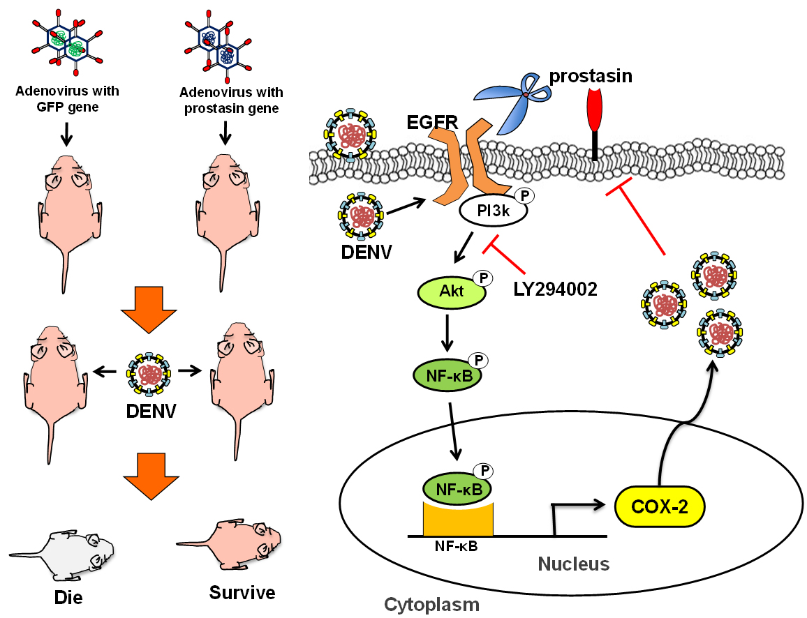
Model for the mechanism of prostasin-mediated inhibition of DENV replication via modulation of EGFR proteolysis.
Exogenous expression of prostasin protected ICR suckling mice from DENV-induced mortality and suppressed DENV propagation. The antiviral mechanism of prostasin was the reduction effect on EGFR, leading to suppression of the Akt/NF-kB-mediated cyclooxygenase 2 (COX-2) signaling pathway.
This article-“Prostasin Impairs Epithelial Growth Factor Receptor Activation to Suppress Dengue Virus Propagation” , written by Rept. Author Prof. Jin-Ching Lee, from Department of Biotechnology, is award for Kaohsiung Medical University 2019 Monthly Excellent Paper Award in May.
Main researcher Introduction:
The team leader is Prof. Jin-Ching Lee. The collaborative clinical leader is Dr. Yen-Hsu Chen (Ph.D.). The co-authors include Chun-Kuang Lin as first author (a Ph.D. student), Chin-Kai Tseng, Yu-Hsuan Wu, Chun-Yu Lin (a clinical Doctor), Chung-Hao Huang (a clinical Doctor), Dr. Weng-Hung Wang, Prof. Chih-Chuang Liaw.
Author Email
Prof. Jin-Ching Lee: jclee@kmu.edu.tw
Dr. Yen-Hsu Chen: infchen@gmail.com
Paper cited from:
This study was published online in The Journal of Infectious Diseases, the 16th of April 2019. The full research article entitled “Prostasin impairs epithelial growth factor activation to suppress dengue virus propagation”
Paper online website: https://academic.oup.com/jid/article-abstract/219/9/1377/5200723?redirectedFrom=fulltext
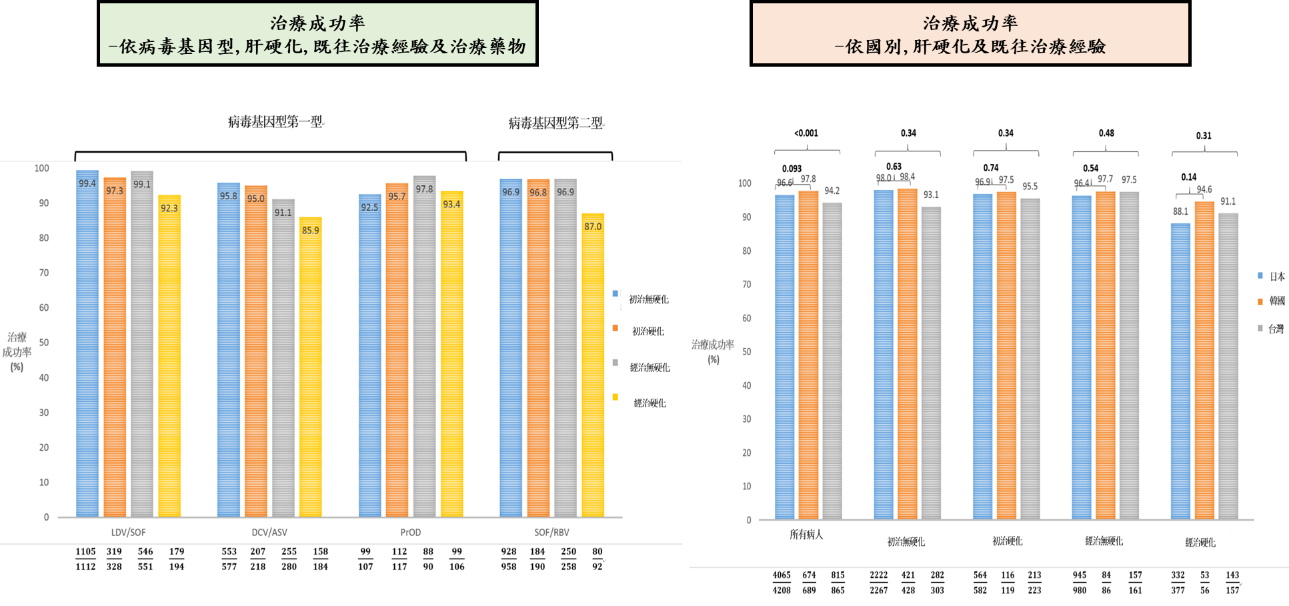
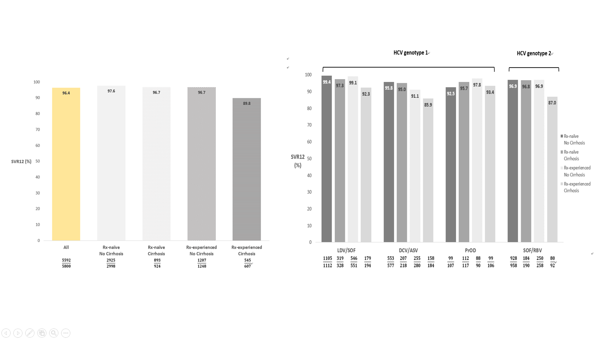
全球有1/3的C型肝炎患者位於亞洲,慢性C型肝炎小分子抗病毒藥物(DAA)既往亞洲真實世界報告多為日本單中心或單一醫療體系發表。亞洲C型肝炎分布人口學及地域分佈複雜,在亞洲多國綜觀的大規模治療報告仍付之闕如。此研究針對亞洲多國慢性C型肝炎患者接受小分子抗病毒藥物的真實世界療效及安全性作進一步探討。REAL-C (Real-world Evidence from the Asia Liver consortium for HCV)為亞洲多國包含台灣、日本、韓國及香港超過20個多中心大規模C型肝炎研究聯盟註冊系統(Registry),此研究的主要目的為探討亞洲C型肝炎族群小分子抗病毒藥物療效(持續性病毒反應;定義為抗病藥物停藥後12週體內偵測不到病毒;SVR12)及安全性研究,以及探討新藥治療失敗原因。本研究收錄來自台灣、日本、韓國及香港一共6,287位慢性C型肝炎患者接受小分子抗病毒藥物。相較其他國家患者,日本患者年紀較老、身體質量比少,且有較高比例非肝癌惡性腫瘤。整體亞洲族群C肝治療療效為96.4%,且無種族之間差別。不同藥物及不同肝病嚴重程度整體治療成果在91.1%至99.4%之間,唯病毒基因型第一型肝硬化干擾素治療失敗患者使用daclatasvir/asunaprevir(87%)及第二型肝硬化干擾素治療失敗患者使用sofosbuvir/ribavirin(85.9%)有較差治療療效。整體治療中斷率為1.9%且無種族之間差別。多變項結果顯示與治療失敗有關因素為高病毒量、肝硬化及之前DAA治療失敗經驗。種族地域差異非影響治療療效之獨立預測因子。此多國多中心研究顯示小分子抗病毒藥物可有效及安全治療亞洲慢性C型肝炎患者,此實證醫學證據可對於疾病負擔沉重的亞洲國家提供C肝治療真實世界證據及各國C肝公衛政策制定之參考。
圖形摘要
本篇為高雄醫學大學2019年月傑出論文9月份得獎研究,代表作者為肝炎研究中心黃釧峰教授。
本校主要研究者之簡介:
黃釧峰 教授
高醫 肝膽內科
高醫大內科學科
研究聯繫Email:
fengcheerup@gmail.com
期刊出處:
Hepatol Int. 2019 Sep;13(5):587-598.
研究全文下載:
https://link.springer.com/article/10.1007/s12072-019-09974-z
Direct-acting antivirals in East Asian hepatitis C patients: Real-world experience from the REAL-C Consortium
Background & Aims
One-third of the global hepatitis C virus (HCV) burden is found in Asia. Real-world data from diverse East Asian cohorts remain limited. This study addressed the real-world status of direct-acting antiviral (DAA) therapy among patients from East Asia.
Methods
Chronic hepatitis C (CHC) patients from clinical sites in Japan, Taiwan, South Korea and Hong Kong were recruited in the REAL-C registry, an observational chart review registry. The primary outcome was sustained virologic response (SVR12, HCV RNA PCR .
Results
A total of 6,287 CHC patients were enrolled. Compared to other East Asian patients, patients from Japan were older (66.3 vs. 61.5 years, p<0.0001), had lower body mass indices (22.9 kg/m2 vs. 24.6 kg/m2, p<0.001), and were more likely to have non-liver malignancy history (12.2% vs. 5.0%, p<0.001).The overall SVR12 rate was 96.4%, similar to patients both inside and outside Japan (96.6% vs. 96%, p=0.21). The SVR12 rate ranged from 91.1-99.4% except treatment-experienced cirrhotic HCV genotype-1 patients who received daclatasvir/asunaprevir (85.9%) and the treatment-experienced cirrhotic HCV genotype-2 patients treated with sofosbuvir/ribavirin (87%). The overall rate of drug discontinuation was 1.9%, also similar across regions. Logistic regression analysis revealed that significant independent factors predictive of treatment failure were higher HCV RNA levels (odds ratio [OR], 95%CI: 0.73, 0.59-0.90; p=0.004), the presence of liver cirrhosis (0.68, 0.49-0.84, p=0.046), prior treatment failure with IFN (0.57, 0.42-0.79, p=0.001), and prior treatment failure with DAA other than BOC and TVR (0.04, 0.02-0.08, p<0.001), but not geographic region (non-Japan versus Japan, 1.09, 0.74-1.60, P=0.68).
Conclusions
In this large multinational CHC cohort from the East Asia, oral DAAs were highly effective and well-tolerated across the region. Policies should encourage treatment for all CHC patients with DAAs in Asia with its heavy burden of HCV.
Graphical Abstract
This article-“Direct-acting antivirals in East Asian hepatitis C patients: real-world experience from the REAL-C Consortium” , written by Rept. Author researcher Chung-Feng Huang, from Hepatitis Research Center, is award for Kaohsiung Medical University 2019 Monthly Excellent Paper Award in Sept.
Main researcher Intro.
Researcher :
Chung-Feng Huang MD.PhD.
Hepatobiliary Division, Department of Internal Medicine, Kaohsiung Medical University Hospital, Kaohsiung Medical University, Kaohsiung, Taiwan
Author Email
Paper cited from:
Hepatol Int. 2019 Sep;13(5):587-598.
Research Paper available online on website https://link.springer.com/article/10.1007/s12072-019-09974-z
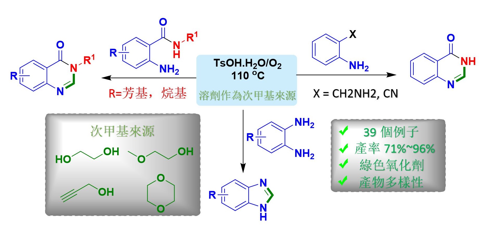
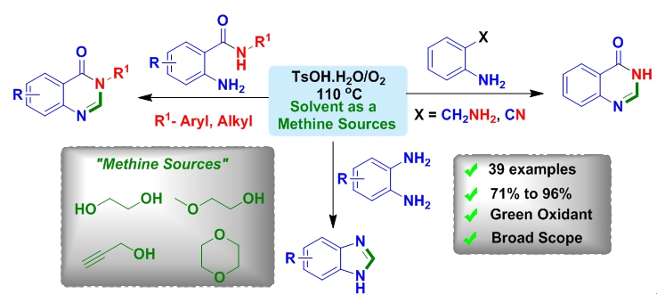
新型環保的次甲基源可作為合成雜環化合物
在最近的研究中,高雄醫學大學的研究人員開發了一種新的化學方法,利用醇類和醚類作為次甲基 –CH 的來源,用於合成喹唑啉酮和苯並咪唑衍生物,這類化合物通常具有藥理特性,如: 抗微生物,抗驚厥,鎮靜,降壓,抗抑鬱,抗炎和抗過敏特性藥物,因此值得推薦應用本方法來合成這類衍生物。
由於前人報導合成喹唑啉的方法受限於產物的範圍、需使用強酸性條件、昂貴的金屬催化劑、爆炸性過氧化物以及長時間反應。 因此,為了克服這些缺陷,王志鉦教授團隊最新研究中發現,在友善環境下以氧氣作為氧化劑,使用醚或醇類為次甲基來源可合成異核芳香族化合物。
此外,這個研究團隊也發現,利用本方法可合成具有生物活性的乙酰膽鹼酮、二咪唑、蕓苔芸香鹼、(±)依夫二胺的前驅物。 隨著乙二醇作為“次甲基”來源的成功,研究團隊進一步確定其他醇和醚亦可作為次甲基來源,此由重氫標記研究證實了這一結果。
這項研究於2019年2月1日發表在《綠色化學》上,名為 ”在無金屬和無過氧化物的條件下,永續次甲基源合成雜環”,可在以下網站在線獲取:https://pubs.rsc.org/en/content/articlepdf/2019/gc/c8gc03839b. (Green Chem., 2019, 21, 979–985)
本論文共同第一作者為Vishal Suresh Kudale和Gopal Chandru Senadi博士,通訊作者為王志鉦教授(高雄醫學大學醫藥暨應用化學系教授 )。
高雄醫學大學醫藥暨應用化學系教授王志鉦教授
電話:+ 886-7-3121101(轉2275),電子郵件:jjwang@kmu.edu.tw
高雄醫學大學醫藥暨應用化學系博士生Vishal Suresh Kudale
電話:+886-7-3121101(轉2275),電子郵件:vishalkudale90@gmail.com
高雄醫學大學醫藥暨應用化學系博士後研究員Gopal Chandru Senadi
電話:+ 886-7-3121101(轉:2275),電子郵件:sgchand@gmail.com
Novel eco-friendly methine sources can synthesize useful heterocycles
In the recent study, the researchers at Kaohsiung Medical University have developed a new chemical method to introduce alcohols and ethers as carbon source (“-CH”) for the synthesis of quinazolinone and benzimidazole derivatives which are highly recommended due to their pharmacological properties, e.g. anti-microbial, anticonvulsant, sedative, hypotensive, anti-depressant, antiinflammatory, and anti-allergy properties.
Previous approaches for the synthesis quinazolines have suffered from limited substrate scopes and strong acidic conditions, expensive metal catalysts, explosive peroxides and long reaction times. So to overcome these drawbacks, in the latest study, Prof. Jeh-Jeng Wang and co-workers found that ethers and alcohols can be used as carbon synthon for the synthesis of heteroarenes in the presence of environmentally benign O2 as an oxidant.
Furthermore, Prof. Wang’s group demonstrated the applicability of this method by synthesizing biologically active echinozolinone, dimedazole, and common precursor of rutaecarpine and (±) evodiamine. With the success of ethylene glycol as a “methine” source, the researchers also identified other alcohols and ethers as a carbon synthons for the synthesis of quinazolinone derivatives. This result was confirmed by Deuterium labelling studies.
This study was published online in Green Chemistry on 1st February 2019 as a research communication entitled “Sustainable methine sources for the synthesis of heterocycles under metal- and peroxide-free conditions” and is available online at https://pubs.rsc.org/en/content/articlepdf/2019/gc/c8gc03839b(Green Chem., 2019, 21, 979–985)
The lead authors of the work entitled “Sustainable methine sources for the synthesis of heterocycles under metal- and peroxide-free conditions” are Vishal Suresh Kudale and Gopal Chandru Senadi, corresponding author include Dr. Jeh-Jeng Wang (Professor, Kaohsiung Medical University, Department of Medicinal and Applied Chemistry, Taiwan).
Media Contact:
Prof. Jeh-Jeng Wang, Kaohsiung Medical University, Department of Medicinal and Applied Chemistry Tel: +886-7-3121101(Ext: 2275), E-mail: jjwang@kmu.edu.tw
Vishal Suresh Kudale, Doctoral fellow, Kaohsiung Medical University, Department of Medicinal and Applied Chemistry Tel: +886-7-3121101(Ext: 2275), E-mail: vishalkudale90@gmail.com
Gopal Chandru Senadi, Postdoctoral fellow, Kaohsiung Medical University, Department of Medicinal and Applied Chemistry Tel: +886-7-3121101(Ext: 2275), E-mail: sgchand@gmail.com
版權所有 © 2021-2025 高雄醫學大學
