

紅斑性狼瘡與低密度脂蛋白引起血管老化的關聯
醫技系柯良胤副教授與過敏免疫風濕內科顏正賢教授、血脂生科研究中心詹華蓁博士…等人共同發表的研究論文「Role of low-density lipoprotein in early vascular aging associated with systemic lupus erythematosus」將於2020年6月刊登於Arthritis & Rheumatology期刊 (Jan 28, 2020 Epub),該論文受到Nature Reviews 期刊編輯Joanna Clarke的關注與採訪,並隨即發表於Nature Reviews Rheumatology研究亮點,文章題目為LDL subfraction linked to vascular ageing and heart disease in SLE。
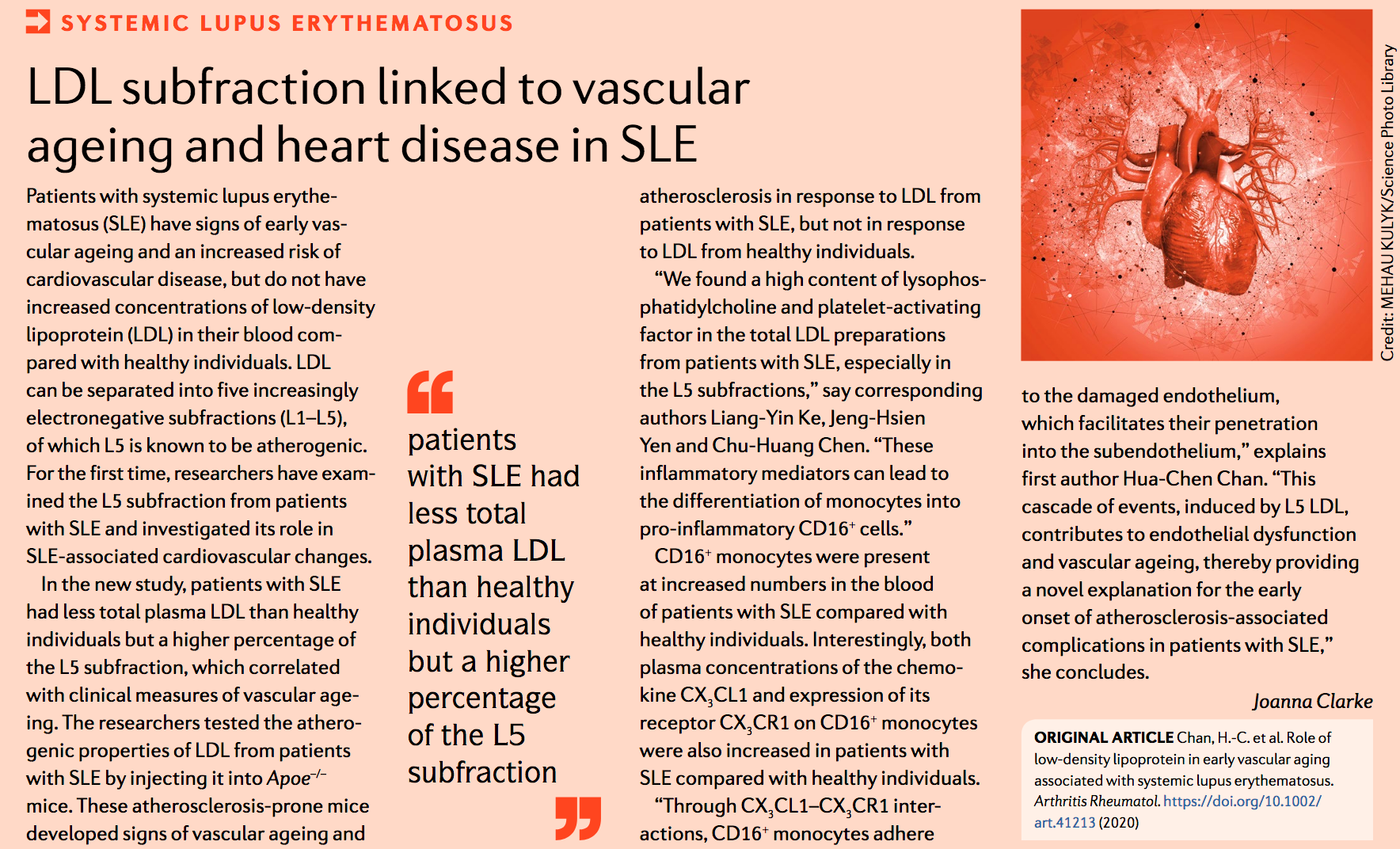
圖一:Nat Rev Rheumatol. 2020 Apr;16(4):187.
陰電性低密度脂蛋白與紅斑性狼瘡病人出現血管過早老化及發生心血管疾病有關
紅斑性狼瘡 (Systemic Lupus Erythematosus; SLE) 病人族群年輕,血液中低密度脂蛋白膽固醇濃度也不高,但卻非常容易發生血管過早老化、動脈粥狀硬化及其他心血管疾病,這是過去一個尚未被透徹研究的臨床重要議題。高醫附院血脂生科研究中心與過敏免疫風濕內科醫師團隊基於過去在高醫校院建立的陰電性低密度脂蛋白 (electronegative low-density lipoprotein; L5 LDL) 特色研究基礎,從而探討SLE病人血液中L5 LDL濃度增加及其引起心血管疾病的致病機轉。
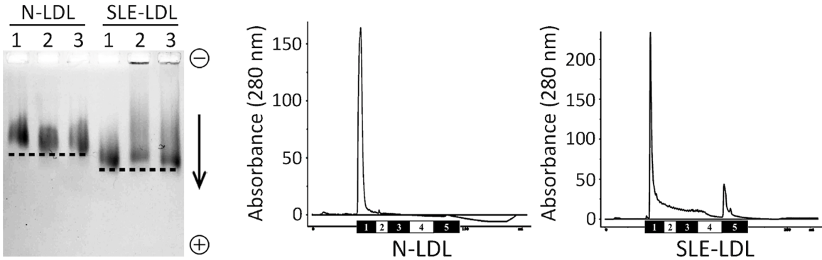
圖二: 以電泳及液相層析法證實SLE病人的LDL陰電性增加
紅斑性狼瘡病人的溶血磷脂質與血小板活化因子濃度增加,尤其增加在L5 LDL次群
LDL的主要特殊成分為高濃度的溶血卵磷脂(lysophosphatidylcholine; LPC) 與血小板活化因子(platelet-activating factor; PAF),這些生物活性的脂質容易造成血管內皮細胞老化與細胞凋亡,也會引起免疫細胞與血小板活化,與心腦血管、內分泌代謝、癌症…等疾病都息息相關。其中,LPC 主要是經由磷脂酶(phospholipase A2)所產生代謝物,需要被快速被帶回肝臟清除;而SLE病人就是代謝產物增加或者來不及清除,因此LPC堆積在血液中而引起一連串的發炎病理反應。
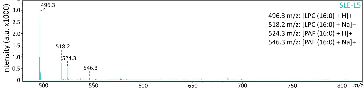 圖三: 以液相層析質譜儀分析SLE病人的LDL得知其主要成分增加了LPC與PAF
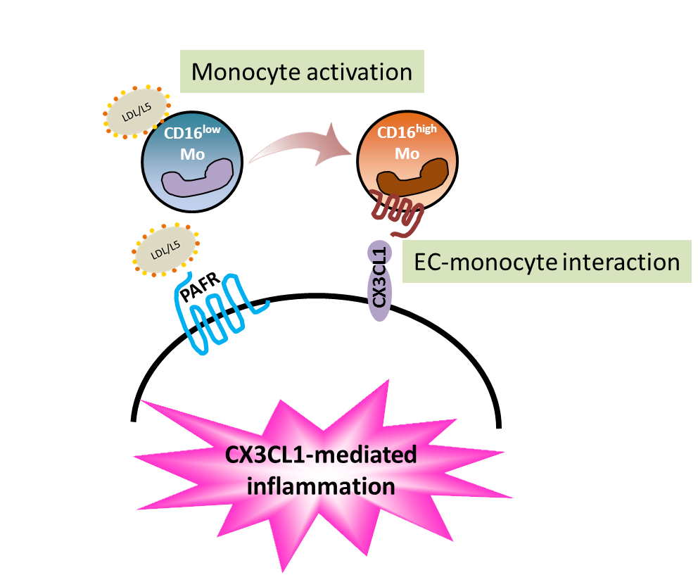
LPC與PAF會增加CD16+單核球細胞,透過CX3CL1-CX3CR1 交互作用造成血管老化
過去已知CD16+單核球細胞帶有CX3CR1細胞接受器,會與受傷的血管內皮細胞CX3CL1+有交互作用。而這項研究發現,LPC與PAF會讓白血球分化、增生為CD16+單核球細胞,同時會破壞血管內皮細胞,使其分泌CX3CL1;透過CX3CL1-CX3CR1 交互作用,讓CD16+單核球浸潤血管內皮層,導致血管功能失調與血管老化,這個病理機制解釋了為什麼SLE病人往往有心血管疾病的病發症,也提供可以改變未來治療策略的見解。
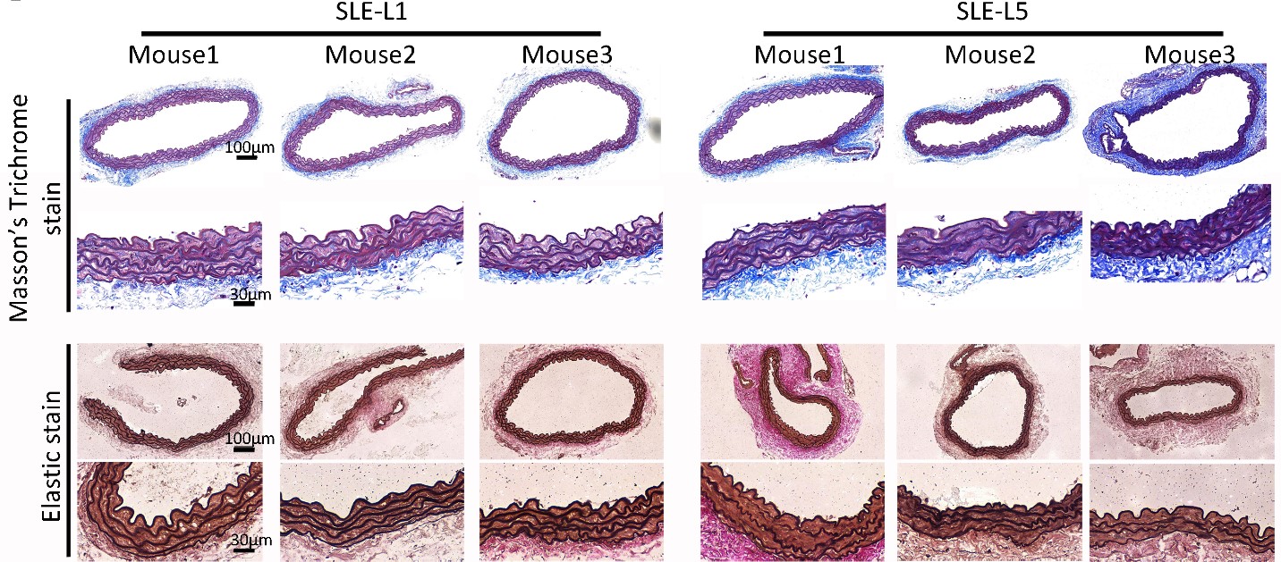
圖四: 注射SLE L5 LDL會加速apoE KO老鼠的主動脈血管老化
結論
紅斑性狼瘡病人血液中陰電性低密度脂蛋白L5 LDL濃度增加,並具有高濃度的生物活性的脂質LPC與PAF,這會引起單核球細胞分化成傾向發炎反應的CD16+細胞,導致血管內皮細胞活化。透過CX3CL1-CX3CR1 分子交互作用使血管功能失調與血管老化。
Graphical abstract
Liang-Yin Ke (from the Department of Medical Laboratory Science and Biotechnology), Jeng-Hsien Yen (from Graduate Institute of Medicine), the first author Hua-Chen Chan (Center for Lipid Biosciences) and colleagues published the above-named paper in the Arthritis & Rheumatology this year. Immediately, the paper got great attention. The editor of Nature Reviews, Joanna Clarke, interviewed with the corresponding authors, and later she wrote a “research highlight” editorial and published in Nature Reviews Rheumatology, entitled as “LDL subfraction linked to vascular aging and heart disease in SLE.”
Figure 1. An research highlight published in Nat Rev Rheumatol. 2020 Apr;16(4):187.
L5 LDL linked to vascular aging in patients with Systemic Lupus Erythematosus (SLE)
SLE patients often have atherosclerotic complications at a young age but normal low-density lipoprotein (LDL) levels. Thus, this study was aimed to explain the paradoxical findings of early vascular aging (EVA) and normolipidemia in patients with SLE. In KMU, we focused on a different approach: the electronegative LDL. Human plasma LDL can be separated into five subfractions. L5 LDL, in particular, has been shown to be atherogenic, independent of LDL cholesterol. From here, we investigated the mechanisms of vascular aging in SLE patients.
Figure 2. SLE patients show elevated levels of atherogenic lipoproteins, L5 LDL.
The mass spectrometric analysis revealed that lysophosphatidylcholine (LPC) and platelet-activating factor (PAF) were increased in SLE-LDL and L5.
LPC and PAF are bioactive lipids, showing the capability to induce inflammatory changes in vascular cells both in vivo and in vitro. In this study, repeated injections of SLE-LDL, LPC or PAF to apoE-/- mice led to increases in IMT, collagen deposition, fatty-streak areas in the aortic wall, and cell senescence in the aortic endothelium.
Figure 3. LPC and PAF were increased in SLE-LDL and L5.
SLE-L5 induces CD16+ expression in monocytes and monocyte adhesion to ECs through CX3CL1-CX3CR1 interactions.
Flow cytometric analysis showed that SLE-L5 lipids, but not SLE-L1 lipids, induced an increase in the percentage of CD16+ monocytes, which are CX3CR1 expressing cells. On the other hand, SLE-LDL induces CX3CL1 expression in activated endothelial cells. Through CX3CL1-CX3CR1 interactions, SLE-L5 induces early atherosclerotic changes in young apoE-/- mice. SLE-L5 induced significant atherosclerotic lesion formation in the aortic arch of the mice as well as collagen deposition and elastic fragmentation in aortas.
Figure 4. : SLE-L5 induced collagen deposition and elastic fragmentation in aortas of mice.
Conclusion. L5 LDL (the most electronegative subfraction) is increased in SLE patients. By mass spectrometry, results showed that L5 LDL is an LPC- and PAF-rich lipoprotein. Through CX3CL1-CX3CR1 interactions between ECs and CD16+ monocytes, L5 LDL promotes atherosclerosis. Our findings suggest that the L5 level, not the total LDL concentration, should be a therapeutic target for preventing EVA and atherosclerosis in patients with SLE.
Graphical abstract