

高醫團隊發現口腔癌新致病機轉及潛在治療方式
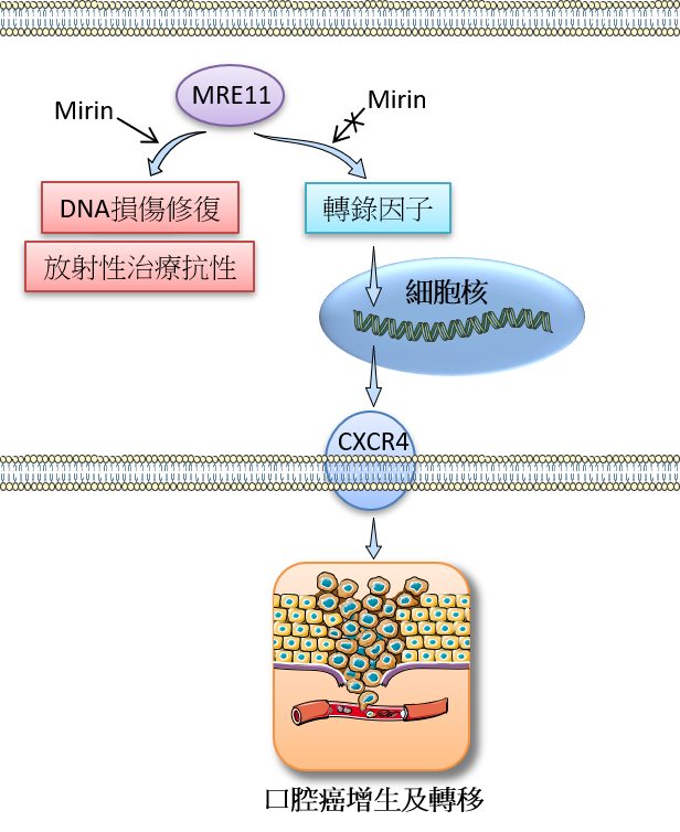
口腔癌在台灣是癌症致死的主要原因之一,尤其好發在台灣男性族群。然而,它的生物機轉及專一性治療策略仍然有待進一步的研究。MRE11是一種與去氧核醣核酸(DNA)的損傷修復有關的重要蛋白,其核酸水解酶活性已知與多種癌症的發展有關連性;然而,MRE11的非核酸水解酶功能卻鮮為人知。在這篇研究報告中,我們新發現MRE11的非核酸水解酶活性與口腔癌的發展有密切相關,並且證實此相關性是透過調控化學趨化受體CXCR4的表現量及其訊息傳遞來完成。
我們首先發現口腔癌病人檢體中的MRE11表現量高低與其腫瘤大小、癌症進展的分期和癌細胞轉移淋巴結之間有正相關,而且MRE11表現量越高的口腔癌病人其預後越差,並易於對放射性治療產生抗性導致治療結果不佳。我們接著用細胞及動物實驗探討這些臨床症狀的生物機轉,發現較多的MRE11會促進CXCR4的表現,並透過其訊息傳遞路徑的活化造成口腔癌細胞的增生及轉移。我們進一步發現這些現象並不是來自MRE11的核酸水解酶活性,而是來自其非核酸水解酶活性,因為當我們加入一種稱為mirin(專門用來抑制MRE11核酸水解酶活性)的抑制劑時,並不會影響經由MRE11所引起的口腔癌細胞增生及轉移的現象。此外,當我們加入CXCR4抗體於細胞或動物實驗時,皆發現此經由MRE11所引起的口腔癌細胞增生及轉移的現象被抑制了。
綜合上述,調控MRE11的表現量將可能成為治療口腔癌的一個新的標的,我們也預期本研究成果能成為臨床診斷口腔癌之重要參考。此外,未來也許可以根據口腔癌病人不同的MRE11表現量發展出個人化的醫療策略。
圖形摘要:
應用與亮點:
§ 口腔癌組織中高表現量的MRE11與口腔癌惡化指標以及病人接受放射性治療後的預後不佳現象具有密切關聯
§ MRE11透過上調化學趨化受體CXCR4的表現與其訊息傳遞來促進口腔癌細胞增生和轉移的能力
§ MRE11的非核酸水解酶功能是促進上述口腔癌細胞增生和轉移現象的主因
§ 預期MRE11可應用在臨床上成為口腔癌預後的重要標記與新的治療標的
【研究團隊】
汪硯雲副教授和袁行修教授的研究團隊長期致力於口腔癌之病因、致病機轉、治療及預防的研究及服務,團隊成員包括汪硯雲、陳玉昆、Steven Lo、紀宗辰、陳怡華、胡楚松、陳雅雯、江士昇、蔡芳瑜、劉旺達、李瑞年、謝雅晶、黃志仁、袁行修。
代表單位:
高雄醫學大學口腔醫學院牙醫學系及醫學院醫學研究所
團隊主要成員簡介:
1. 汪硯雲博士是高雄醫學大學口腔醫學院牙醫學系專任副教授,同時也是高雄醫學大學癌症研究中心及高醫附院轉譯醫學研究中心的成員。
研究聯繫Email:wyy@kmu.edu.tw
2. 袁行修博士是高雄醫學大學醫學院醫學研究所專任教授,同時也是高雄醫學大學癌症研究中心及高醫附院轉譯醫學研究中心的成員。
研究聯繫Email:yuanssf@kmu.edu.tw
【論文資訊】
論文出處:Oncogene. 2021, 40: 3510–3532.
全文下載:https://www.nature.com/articles/s41388-021-01698-5
New pathogenic mechanism and potential treatment of oral cancer
Oral
cancer is one of the leading cause of cancer deaths in Taiwan, prevalently
occurring in the male population. However, its biological mechanisms and
specific therapeutic strategies remain to be explored. MRE11 is the core
component of the RAD50/MRE11/NBS1 complex for repair of DNA
double-strand-breaks, and has been known as a critical factor in solid tumor
development through its nuclease-dependent activity. In this article, we found a
novel role of MRE11 in oral cancer progression by its nuclease-independent function,
and delineated its key downstream signaling through the chemoattractant
receptor CXCR4 as follows. First, MRE11 expression in oral cancer samples was
positively associated with the tumor size, cancer stage, and lymph node
metastasis, and was predictive of poorer patient survival and radiotherapy
resistance. In addition, MRE11 promoted oral cancer cell proliferation and migration/invasion
ability, and the cancer-promoting effects of MRE11 were mediated by enhancing CXCR4
expression and its signaling. Notably, these phenomena were mitigated by using CXCR4
neutralizing antibodies in both in vitro
and in vivo models. Moreover,
inhibition of the nuclease-dependent activity of MRE11 by mirin reduced DNA
repair of oral cancer cells; however, mirin treatment did not affect
MRE11-promoted oral cancer cell proliferation and migration/invasion ability as
described above, indicating that the nuclease-independent function of MRE11
underlies these pro-cancer effects. Collectively, MRE11 may serve as a clinical
indicator and therapeutic target in oral cancer, displaying dual nuclease-dependent
and independent roles that permit separate targeting of tumor vulnerabilities
in oral cancer treatment. These findings will also facilitate future
development of personalized medicine according to the differential expression
of MRE11 in oral cancer patients.
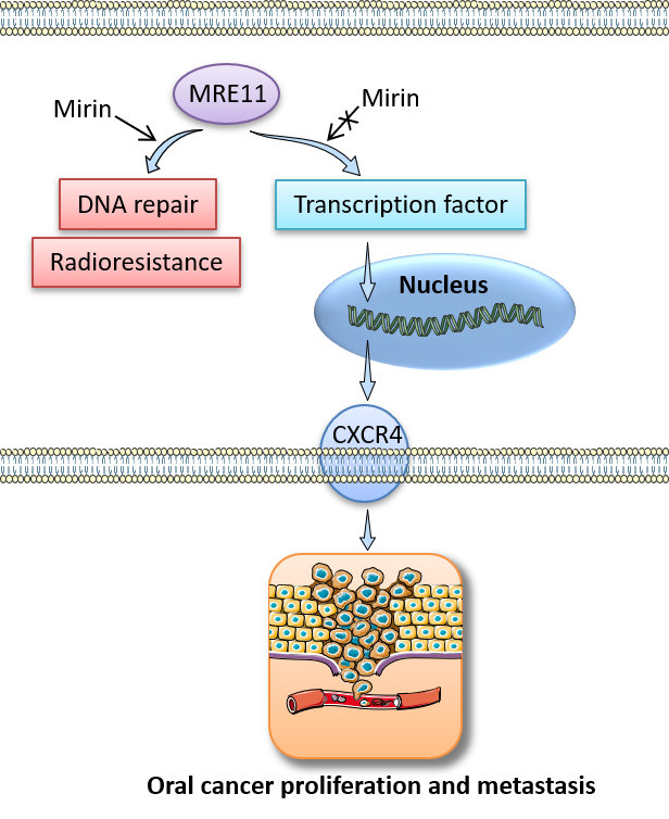
Graphical
Abstract:
Application
and Highlights:
§ High expression level of MRE11 in oral
cancer tissues is associated with adverse oral cancer progression and treatment
outcomes by radiotherapy
§ MRE11 promotes oral cancer cell
proliferation and migration/invasion ability through upregulation of
chemoattractant receptor CXCR4 and its signaling
§ These cancer-promoting effects result
from the nuclease-independent function of MRE11
§ MRE11 may have clinical benefits as a
prognostic indicator and novel target for oral cancer treatment
Research
Team Members:
Associate Professor Yen-Yun Wang and Professor Shyng-Shiou F. Yuan are dedicated to the translational research
of the etiology, pathogenesis, treatment and prevention of oral cancer. Team
members including Yen-Yun Wang, Yuk-Kwan Chen, Steven Lo, Tsung-Chen Chi, Yi-Hua Chen, Stephen Chu-Sung Hu, Ya-Wen
Chen, Shih Sheng Jiang, Fang-Yu Tsai, Wangta
Liu, Ruei-Nian
Li, Ya-Ching
Hsieh, Chih-Jen
Huang & Shyng-Shiou
F. Yuan
Representative Department:
Associate Professor Yen-Yun
Wang:
School
of Dentistry, College of Dental Medicine, Kaohsiung Medical University, Kaohsiung,
Taiwan
Professor Shyng-Shiou F. Yuan:
Graduate
Institute of Medicine, College of Medicine, Kaohsiung Medical University
Introduction
of Research Team:
The
first author Yen-Yun Wang is a full-time associate professor in the School of
Dentistry, Kaohsiung Medical University, and an appointed member of the Center
for Cancer Research, Kaohsiung Medical University and Translational Research
Center, Kaohsiung Medical University Hospital.
The
corresponding author Shyng-Shiou F. Yuan is a full-time professor in the Graduate
Institute of Medicine, Kaohsiung Medical University, and an appointed member of
the Center for Cancer Research, Kaohsiung Medical University and Translational
Research Center, Kaohsiung Medical University Hospital.
Contact
Email:
Associate
Professor Yen-Yun Wang: wyy@kmu.edu.tw
Professor
Shyng-Shiou F. Yuan: yuanssf@kmu.edu.tw
Publication:
Oncogene.
2021, 40: 3510–3532.
Full-Text
Article: https://www.nature.com/articles/s41388-021-01698-5
建立快速螢光檢測平台應用於診斷人體基因變異
近幾年來,大眾開始對健康與預防醫學的重視,而使得【基因檢測】成為相當重視的國家發展主題與學術研究項目。自2000年人類基因體計畫完成以來,越來越多的基因功能被解讀成功,已超過2000種基因相關疾病被發現,如今已有700多種基因相關疾病已開發出相應藥物及治療方法,且癌症與許多罕見疾病。基因檢測是偵測染色體結構中之DNA序列是否發生變異位點或基因表現程度,提供受檢者與醫療研究人員評估一些與基因遺傳有關的疾病、體質或個人特質的依據,也是精準醫學分析的一種方法。每一個人的DNA基因都是獨特的個人化資訊,造成每一個人的先天體質,健康狀況,特徵都不相同;2008年,美國時代雜誌曾經把這個革命性技術評選為2008年度最佳創新之首。
而現行基因檢測技術經常需要使用到一些昂貴耗材或大型儀器,使得檢測費用較高 (約2000-5000元等),且須時數日至數週,因此並非所有實驗室皆可完成之檢驗技術,而為了使基因檢測技術更簡單、方便、準確且快速,本研究開發一『快速應用螢光於基因變異檢測之技術』,應用於分析運動神經元存活基因(survival motor neuron gene,簡稱SMN)以診斷脊髓肌肉萎縮症(spinal muscular atrophy,簡稱SMA)。
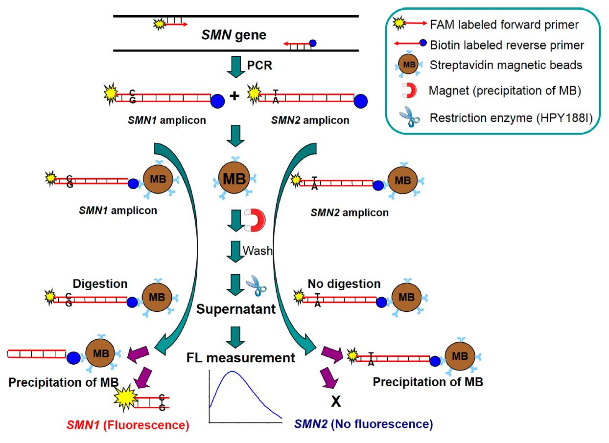
脊髓肌肉萎縮症為一種先天遺傳的疾病,造成病患肌肉無力且萎縮,輕症者行動不便,重症者則會因呼吸衰竭而死亡。現行已知運動神經元存活基因為控制此疾病之基因,於懷孕初期診斷該基因可96%預防此疾病發生或進行即早治療,因此該基因檢測已是懷孕婦女必檢測之基因項目之一。目前為止,臨床上有數種方法可用來診斷該基因,包括有限制酶酵素分解法(RFLP),real-time PCR方法、denatured HPLC (DHPLC),毛細管電泳法、質譜分析法或Multiplex Ligation-dependent Probe Amplification (MLPA)等,但該些方法就如同前述的傳統基因分析方法,分析耗時且需要一些昂貴耗材或大型儀器。而本研究為使基因檢測更加簡單且快速,設計一快速螢光基因檢測平台,其原理如圖形摘要所示,僅須於引子對修飾Biotin基團與螢光基團,另搭配磁珠基因捕捉與純化,最後用限制酶酵素將捕捉基因片段進行剪切,即可應用螢光的強弱變化,了解該運動神經元存活基因為何種基因型。此方法方便、簡單且快速,且該方法具有通用性,只要選擇適當的限制酶酵素即可應用於不同基因之檢測,相當地適合應用於分析各種不同類型的基因變異。
圖形摘要
應用與亮點:
1. 基因檢測是偵測染色體結構中之DNA序列是否發生變異位點或基因表現程度,提供受檢者與醫療研究人員評估一些與基因遺傳有關的疾病、體質或個人特質的依據,也是精準醫學分析的一種方法。
2. 本研究開發一『快速應用螢光於基因變異檢測之技術』,應用單純螢光強弱於分析運動神經元存活基因(survival motor neuron gene,簡稱SMN)以診斷脊髓肌肉萎縮症(spinal muscular atrophy,簡稱SMA)。
3. 此方法方便、簡單且快速,且該方法具有通用性,只要選擇適當的限制酶酵素即可應用於不同基因之檢測,相當地適合應用於分析各種不同類型的基因變異。
【研究團隊】
團隊成員:王俊棋、陳俊安、鐘育志、柯黃盛
代表單位:高雄醫學大學 藥學系
團隊簡介:
該研究團隊長年來致力於毛細管電泳法、藥物分析、基因分析、特殊探針及引子設計、奈米粒子合成及應用等,已開發出多項藥物監測或基因檢測之技術,且獲得多項專利。
研究聯繫Email:chunchi0716@kmu.edu.tw
【論文資訊】
論文出處:Analytical Chemistry 2018, 90(19): 11599–11606.
全文下載:https://pubs.acs.org/doi/10.1021/acs.analchem.8b02996
Development of Fast Fluorescent Genotyping Platform for Detection of Nucleotide Variants in Human Genes.
In recent years, the public has begun to attach importance to health and preventive medicine, which has made genetic testing become a national development theme and academic research project. Since the completion of the Human Genome Project in 2000, more and more gene functions have been successfully interpreted, and more than 2,000 gene-related diseases have been discovered. Today, more than 700 gene-related diseases have been developed with corresponding drugs, treatments, cancer and many rare diseases. Genetic testing is to detect whether the mutation sites of the DNA sequence in the chromosome structure or the degree of gene expression, which provides the information for medical researchers to evaluate some diseases, physiques or personal characteristics related to genetic inheritance. Genetic testing is also a part of the precision medicine. Everyone's DNA genes are unique and personalized information, which causes everyone's innate physique, health status, and characteristics. In 2008, the American Time magazine selected this revolutionary technology as the top innovation of 2008.
The current genetic testing technology often requires the use of some expensive consumables or large-scale instruments, which makes the testing cost high, and takes several days to several weeks. Therefore, not all laboratories can complete the testing technology. In order to make the gene detection technology simpler, more convenient, accurate and fast. This research has developed a technique for rapid application of fluorescence in gene mutation detection, which is applied to analyze survival motor neuron gene (SMN) for diagnosis of spinal muscular atrophy (SMA).
SMA is a congenital genetic disease that causes muscle weakness and atrophy in patients. People with mild disease have inconvenience in mobility, and those with severe disease will die due to respiratory failure. The SMN gene that controls this disease has been known. Diagnosing this gene in the early stages of pregnancy can prevent the disease from occurring 96% or provide immediate treatment. Therefore, this gene testing is one of the genetic items that must be tested for pregnant women. So far, there are several clinical methods for diagnosing this gene, including restriction enzyme digestion (RFLP), real-time PCR, denatured HPLC (DHPLC), capillary electrophoresis, mass spectrometry or multiplex ligation-dependent probe amplification (MLPA), etc., but these methods are just like the aforementioned traditional genetic analysis methods. These analyses are time-consuming and require some expensive consumables or large instruments. In order to make gene detection simpler and faster, this study designed a fast fluorescent gene detection platform. The principle is as shown in the summary. Only the primer pair needs to modify with the biotin group and the fluorescent group, respectively. This gene amplification was captured with magnetic beads, and finally cut with restriction enzymes. The fluorescent intensity changes can be used to understand the genotype of the survival motor neuron gene. This method is convenient, simple and fast, and the method is universal. As long as the appropriate restriction enzyme is selected, it can be applied to the detection of different genes, and it is quite suitable for the analysis of various types of gene mutations.
Graphical Abstract
Application and Highlights:
1. Genetic testing is to detect whether the mutation sites of the DNA sequence in the chromosome structure or the degree of gene expression, which provides the information for medical researchers to evaluate some diseases, physiques or personal characteristics related to genetic inheritance. Genetic testing is also a part of the precision medicine.
2. In order to make the genetic testing technology simple, convenient, accurate and fast, this research develops a fast fluorescent detection technology in the analysis of gene mutations for determination of survival motor neuron gene (SMN) to diagnose spinal muscular atrophy (SMA).
3. This method is convenient, simple, fast, and versatile. It can be applied to the detection of different genes as long as the appropriate restriction enzymes are selected, and it is quite suitable for the analysis of various types of gene mutations.
Research Team Members:
Chun-Chi Wang, Chung-An Chen, Yuh-Jyh Jong, Hwang-Shang Kou
Representative Department:
School of Pharmacy, College of Pharmacy, Kaohsiung Medical University
Introduction of Research Team:
The team has been committed to capillary electrophoresis, pharmaceutical analysis, genetic analysis, special probe and primer design, nanoparticle synthesis and application, etc. for many years. They have developed a number of technologies for drug monitoring or genetic testing, and obtained a number of patents.
Contact Email: chunchi0716@kmu.edu.tw
Publication: Analytical Chemistry 2018, 90(19): 11599–11606.
Full-TextArticle: https://pubs.acs.org/doi/10.1021/acs.analchem.8b02996
版權所有 © 2021-2025 高雄醫學大學
